The deep intrinsic muscles are located beneath the erector spinae, and are known collectively as the transversospinales. They are a group of short muscles, associated with the transverse and spinous processes of the vertebral column that primarily support the segments of the spine and serve as proprioceptors. Personal trainers who want to go deeper into their anatomy knowledge and application should read on.
The Many-Layered Muscles of the Back
Understanding the complexities of the muscles of the back can be simplified by first grasping that all the muscles of the back comprise one of three layers:
- Superficial (the larger movers of the shoulder and scapulae such as the latissimus dorsi, traps and rhomboids)
- Intermediate (the muscles of the posterior thorax that assist in breathing: Serratus posterior superior and inferior)
- Deep (the deep, intrinsic muscles that support the spine)
The three layers of the deep, intrinsic muscles are categorized into the same designations listed above, which can make matters confusing:
- Superficial Intrinsic (the spinotransversales): The top layer of intrinsic muscles of the cervical spine.
- Intermediate Intrinsic (the erector spinae): The middle layer that extends from the lumbosacral fascia up to the cranium
- Deep Intrinsic (the transverospinales): The deepest layer of tiny local stabilizer muscles that primarily control the segmental motions of the spine. (the subject of this blog)
We’ve explored the first two layers of the intrinsic muscles of the back: The Spinotransversales—the most superficial layer of vertebral column musculature— and the Erector Spinae group—the intermediate layer of muscles supporting the spine.
Three Groups of the Deep Intrinsic Back Muscles (plus a bonus group)
The deeper layer of intrinsic muscles of the back are more propioceptive in function rather than likely to perform agonist actions that the more superficial muscles are responsible for. That is, they provide information to the more global structures about flexion, extension, and rotation, while also performing vital stability to the segments of the spine.
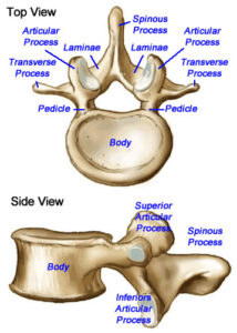
(Familiarity of the vertebral landmarks will help understand where the following muscles attach)
Semispinalis: Not to be confused with the spinalis muscle of the erector spinae group, the semispinalis is located more lateral to the vertabral column and its function is to extend the cervical spine and contralaterally rotate the head and vertabral column. It is divided into two segments: capitis and cervicis.
- Semispinalis capitis: (inserts from the transverse processes of C7-T6 coursing superomedially (up and center) to insert between the superior and inferior nuchal lines of the skull).
- Semispinalis cervicis: (inserts on the transverse processes of T6-T12 into the spinous processes of C1-C5).

Multifidus: Inferior to semispinalis and running from the sacrum to the cervical spine, this muscle has most influence in the lumbo-sacral region and provides stability to the vertebral column.
It has muscle insertions from the sacrum, iliac crest and erector spinae aponeurosis (the broad, sheet-like fascia of the erector spinae) and into the spinous processes of each vertebra. It has attachements in the transverse processes of T1-T3 and articular processes of C4-C7. Each fiber ascends between 2 and 4 vertebral segments, connecting the spinous processes of the vertebrae.
- Multifidus thoracis: (attaches from the transverse processes of the thoracic vertebrae coursing superomedial course to insert variably on the spinous processes of the vertebrae 2 – 5 levels above)
- Multifidus lumborum: (arises from the mammillary processes of the lumbar vertebrae and the posterior surface of the sacrum, PSIS and the posterior sacroiliac ligament coursing superiorally to insert on the spinous processes about 2 to 5 levels above their origin).
While the multifidus has the capability of extending, rotating and laterally flexing the segments of the spine, it is very weak in these areas and functions more as a stabilizer.
- Rotatores brevis: Crosses only one vertrebra to attach to the spinous process to the vertebra superior to it.
- Rotatores longis: Crosses two vertebrae, attaching to the spinous process to the vertebra two levels above.
Minor Deep Intrinsic Muscles:
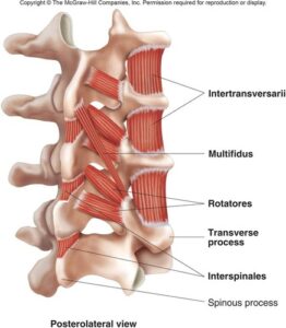
Look closely at the photo above. Bear in mind you are looking at the spine from a posterior and slightly lateral perspective. It becomes very clear that the role of these tiny muscles is not agonistic, but rather to stabilize.
- Interspinales: Connects adjacent spinous processes, as the name suggests.
- Intertranversari: Connects adjacent transverse processes, as the name suggests!
- Levatores costarum: Inserts from the transverse processes of C7-T11 to the inferior rib and performs rib elevation and assists in thoracic rotation in addition to providing vertebral stability.
Why Does a Personal Trainer Need to Know All This?
Well, if you read this far, congratulations! You must really love anatomy and should most definitely pursue our Anatomy Fundamentals Course. But, seriously, folks….The short answer is, you don’t need to know this backward and forward. Most of the programming you create for your clients will likely not ever involve semispinalis growth.
However, #themoreyouknow…
The greater your understanding of the complicated human machine, the more expert your skillset will be. You may have clients coming out of PT for back troubles where it might help to know where the multifidus is located and what its function is, because they were just informed that this muscle is atrophied after pregnancy and is now not performing its function of stabilizing the lumbosacral segments and causing pain. (Ok, I’m just talking about me now)
This is not an unlikely scenario, however. Nor is a client presenting with terrible spinal posture from a desk job or constant gardening. Knowing how the deep muscles of the back coordinate with each other and the more global muscles will bring you that much closer to identifying the problems you see and helping your client correct them with exercise.

References
kidport.com/RefLib/Science/HumanBody/SkeletalSystem/Vertebrae.htm
teachmeanatomy.info/back/muscles/intrinsic/
geekymedics.com/deep-back-muscles/
kenhub.com/en/library/anatomy/intrinsic-back-muscles